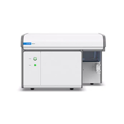
Flow cytometer
NovoCyte Flow Cytometer Systems 1-3 Lasers
본문
NovoCyte is a high-performance benchtop flow cytometer designed for all levels of users and all types of laboratories. The budget-friendly instrument can detect up to 17 parameters with enhanced sensitivity and resolution. The customizable laser and optical configurations of the NovoCyte offer a high degree of flexibility while providing complex cell analysis capabilities.
The NovoExpress software facilitates easy and intuitive sample acquisition and analysis. Automation of multiple fluidic functions eliminates cumbersome and time consuming procedures. Manual sample handling is minimized by the flexible NovoSampler Pro. The system can automatically analyze samples in single tubes, multi-tube racks, or 24-, 48-, or 96-well plates
Features
- Powerful capabilities with up to 17 parameter detection with enhanced sensitivity and resolution.
- Intuitive, advanced data analysis capabilities for greater usability and automated instrument maintenance functions
- Customizable sampling with 1 to 3 laser options, exchangeable filters, multiple sampling options, and flexible analysis formats
- Enhanced usability with fixed optical alignment, 24-bit detection dynamic ranges and no need for PMT voltage adjustment, accurate volumetric-based cell counting, pressure sensors to monitor fluidic status in real-time, and automatic cleaning and decontamination processes
- NovoExpress software is intuitive and easy to use. Flexible analysis templates and plotting tools save time and increase efficiency in data analysis
Specification
| Fluorescence Channels |
|
| Laser Configuration |
|
| Number of Lasers |
|
How it work
The Ultimate Photodetector
Silicon photomultipliers (SiPM) are solid-state, silicon-substrate-based, photon-level-sensitive semiconductor devices, with a 7.2 log dynamic range. Consisting of a compact array of avalanche photodiodes operating in unison, the SiPM is a compact detector with photon counting capability. The innovative optics designed into the NovoCyte Advanteon incorporates 21 independent SiPM, which collect and process signals for each of its fluorescence channels.

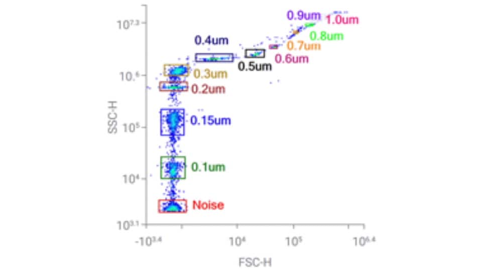
Excellent scatter resolution to detect small particles
Consistent results, fast or slow

Applications
Cytokine Detection
Cytokines are small molecules essential for immune cell response to activation by pathogens, autoimmunity, or therapeutics. Signaling by cytokines can modulate gene regulation, innate and adaptive immune response, and inflammation. Measuring cytokine production and identifying the source of cytokine production is therefore important for an in-depth understanding of the immune response. Bead-based flow cytometry-based detection of cytokines is a highly effective method for measuring multiple soluble analytes in a single sample, using a mixture of bead populations with varying fluorescence intensities.
Intracellular Protein Detection
Detection and analysis of intracellular proteins allow for additional characterization of cell subpopulations and cellular processes. In order to analyze proteins not located on the cell surface, fixation and permeabilization of the cell is required. However, many phospho-specific antibodies are not compatible with many common detergent-based permeabilization methods used for intracellular staining. Special attention is needed when determining the proper fix/perm method for your phospho-specific antibody. The most common method uses 1.5% paraformaldehyde for fixation followed by 100% methanol for permeabilization. While this method works for many antibodies, please note it may not work for every phospho-specific antibody.
Additionally, identifying various cell populations in a heterogenous sample requires staining for phosphorylated proteins coupled with surface proteins. Special consideration must be given to the sensitivity of these epitopes to fixative, taking precaution to avoid damage to the epitope. Therefore, the sample may require staining for specific surface markers before fixation.
Learn More >

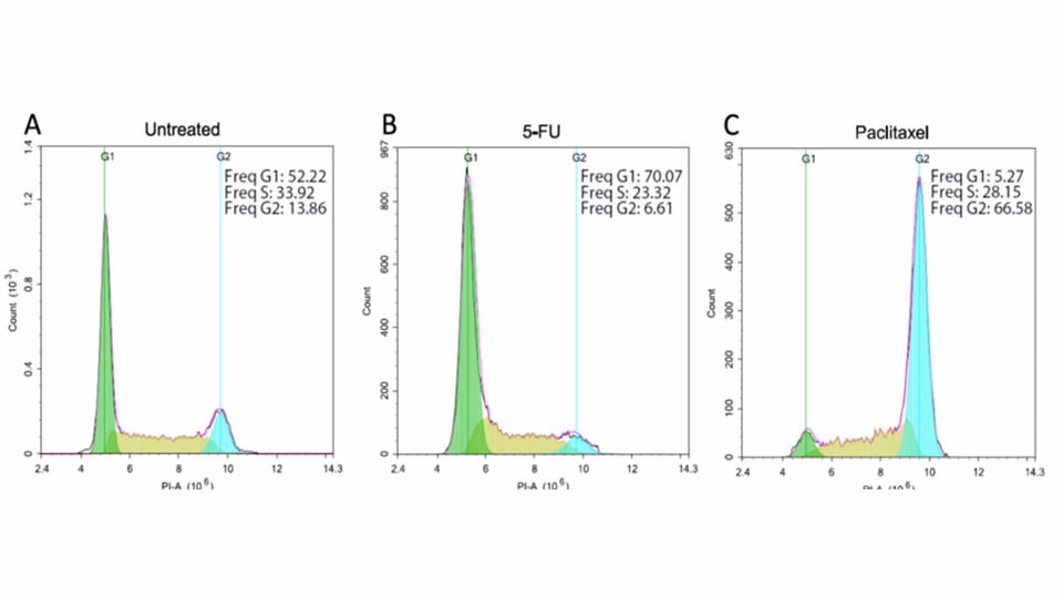
Cell Cycle Analysis
Normal human somatic cells are diploids containing a constant amount of DNA. During cell cycle progression, DNA synthesis results in a doubling of total DNA content, followed by restoration of the normal DNA content after mitosis. Detailed cell cycle analysis can be performed to understand tumor cell differentiation, cell transformation and cell-compound interaction with the NovoCyte flow cytometer.
Figure: After treatment with 10 migrograms/M MG132 or 500 micrograms/M 5-FI for 16 hours/ A549 cells were analyzed for cell cycle distribution with the ACEA Novocyte flow cytometer. The the Novoexpress built-in cell cycle analysis module, the plot shows cells ni G0/G1 phase (green), S phase (yellow) and G2/M phase (blue). Compared to normal untreated cells, MG132 treated cells were arrested at G2/M phase, while 5-FU treated cells were arrested at G0/G1 phase.